
Fast Optical Recording Circuit
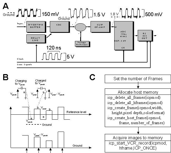
A block diagram of the recording system is presented, which is constructed around an upright microscope mounted on a vibration isolation table. The system includes a CCD detector unit, a digital frame grabber, and a personal computer (PC). A second PC is employed to drive a laboratory interface for synchronizing optical and electrical signals from the cell preparation placed on the microscope stage.
The imaging system described integrates several components to facilitate high-speed imaging in biological research. The CCD sensor, known for its efficiency in low-light conditions, is crucial for capturing images of fluorescently labeled cells. The 41 Msample/s A/D converter ensures that the sensor's output is accurately digitized, maintaining the integrity of the image data. The use of customized electronics allows for optimization of the image acquisition process, enabling rapid frame rates that are essential for observing dynamic cellular processes.
The vibration isolation table is a critical component, as it minimizes external disturbances that could affect image quality, particularly at high magnifications. The dual-PC setup enhances the system's capabilities by allowing one PC to handle image acquisition while the other manages synchronization tasks. This separation of functions ensures that the imaging process runs smoothly and efficiently, providing researchers with reliable data for their experiments.
Overall, the imaging system described is a sophisticated tool designed to meet the demands of modern biological imaging, combining speed, sensitivity, and precision to support advanced research in physiology and neuroscience.This document presents technical details, hardware and software of a complete imaging system which uses a fast CCD sensor and a 41 Msample/s A/D converter to acquire full-frame 12 bit/pixel digitised images with a time resolution of 1.25 ms/image. The noise characteristics and the photon-capture capabilities of the system were analysed in detail. At the maximum readout rate, noise from the electronics amounted to ~300e-, without cooling.
Optical methods are interesting to physiologists and neuroscientists for several reasons. Of particular importance is the capability of recording simultaneously from multiple sites and the possibility to reach fast sampling rates. Therefore it is fundamental to develop imaging systems that, combining both features, achieve a high spatio-temporal resolution.
In this respect, CCD sensors are well suited for detecting low-light level signals from fluorescent probes introduced into living cells. These devices possess high quantum efficiency for converting light into electrical signals that allow monitoring variations in the intracellular concentration of Ca2+ and other fundamental ions within the cell cytoplasm. Here we provide a detailed description of an imaging system based on a sensitive CCD sensor with high readout rate (16 MHz).
Sensor’s output was digitised at 12 bit/pixel by customised electronics permitting image acquisition rates up to 800 frames/s with a full-frame resolution of 128´ 128 pixels.
Fig.1 presents a block-diagram of the recording system that was built around an upright microscope, mounted on a vibration isolation table, and included a CCD detector unit, a digital frame-grabber and a PC. A second PC was used to drive a laboratory interface for the synchronisation of optical and electrical signals from the cell preparation placed on the microscope stage.
🔗 External reference
Related Circuits
This 555 timer is designed uniquely to provide a positive output through control over its reset pin. Typically, the 555 timer IC is triggered by applying a negative voltage. The 555 timer is a versatile integrated circuit widely used in...
This circuit functions as a camera switch, allowing multiple cameras to be connected to a single monitor. It can operate in both manual and automatic modes. In automatic mode, the circuit utilizes a 555 astable multivibrator to generate a...
A street light that automatically switches ON when night falls and turns OFF when the sun rises. The circuit uses an LDR to sense the light. The automatic street light circuit functions by utilizing a Light Dependent Resistor (LDR) as...
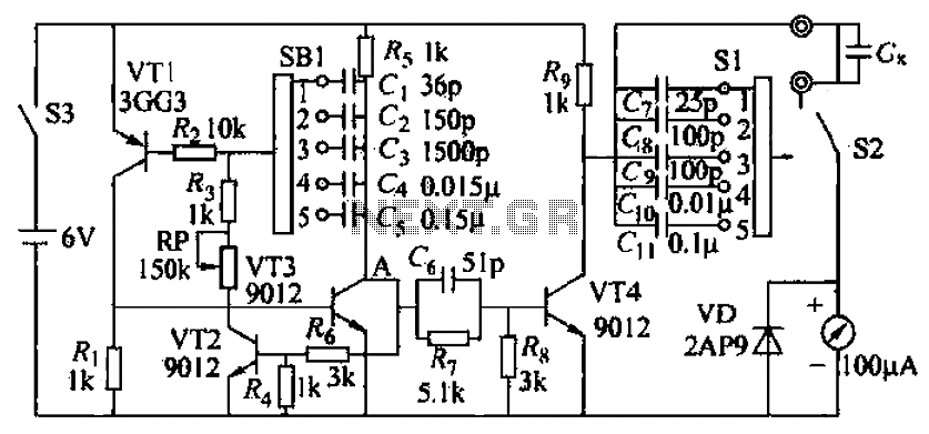
A capacitive measuring instrument is a direct reading device that measures the capacitance of a circuit. This instrument is capable of measuring capacitance values ranging from a few picofarads to 0.1 microfarads, with specific ranges of 25 pF, 100...
Section Ul-a is configured as a high-gain inverting voltage amplifier that is inductively coupled to the phone line via LI. Inductor LI is a homemade unit that consists of 250 turns of fine, enamel-coated wire that is wound on...
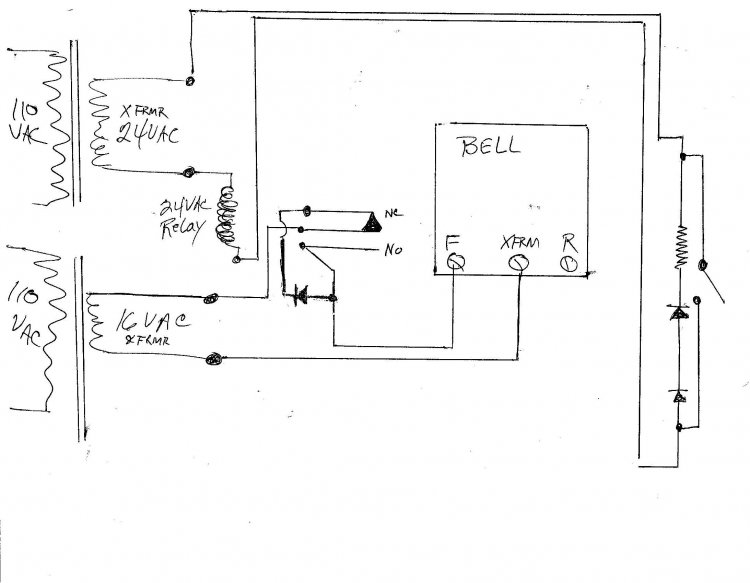
A new wired doorbell has been installed, specifically the Heath/Zenith Model #LE-65-B (Electronic). A new 16 Volt transformer was also added, along with a lighted pushbutton and a diode. Initially, all components functioned correctly, including the lighted pushbutton. However,...
Warning: include(partials/cookie-banner.php): Failed to open stream: Permission denied in /var/www/html/nextgr/view-circuit.php on line 713
Warning: include(): Failed opening 'partials/cookie-banner.php' for inclusion (include_path='.:/usr/share/php') in /var/www/html/nextgr/view-circuit.php on line 713