
magnetic field therapy

The operation of devices that utilize pulsed magnetic fields for microbial destruction is based on well-established principles of electromagnetism and biophysics. The fundamental mechanism involves the generation of ultrasound through oscillating magnetic fields, which interact with charged particles in biological tissues. The oscillations induced by the pulsed magnetic fields create mechanical stress on the protein structures of viruses and bacteria, leading to their disintegration.
The design of such a device typically includes a high-voltage capacitor, a wire coil, and a control circuit to manage the discharge of the capacitor. The wire coil is strategically placed on the surface of the body to maximize the magnetic field's penetration. When the capacitor discharges, it generates a rapidly changing magnetic field, which induces an electric field in the surrounding tissues. This electric field facilitates the oscillation of charged particles, primarily ions, in the body, leading to the generation of ultrasound waves.
The ultrasound frequency generated is proportional to the rate of change of the magnetic field strength. Thus, the design must ensure that the magnetic field can be varied rapidly and maintained at a high intensity. The configuration of the coil and the characteristics of the capacitor play a critical role in optimizing the performance of the device. The coil's geometry can be tailored to enhance the magnetic field distribution and ensure even coverage of the treatment area.
In addition to the ultrasound generation, the transient electric fields produced by the pulsed magnetic fields can induce charge density waves in the electrolytic fluids of the body. These waves propagate through the tissues, creating areas of high and low ion concentration that can further disrupt microbial structures. The dynamics of these charge density waves can lead to the denaturation of proteins on the surfaces of viruses and bacteria, rendering them ineffective.
Overall, the integration of pulsed magnetic fields and ultrasound generation presents a promising approach to microbial destruction. The ability to harness these physical principles in a practical device offers potential applications in medical and therapeutic settings, particularly in combating infections and improving health outcomes. Further research and development are essential to refine these technologies and validate their efficacy in clinical applications.Imagine devices that can disable and destroy microorganisms-viruses, bacteria and fungi - by means of a pulsed, intense, magnetic field! They are not devices for the distant future. They are for today! Already several different pulsed magnetic field instruments are being used in the alternative health field.
Many claims and suggestions are being m ade for and about them. Let`s look into their validity. Let`s also look at a simple device that uses a strong permanent magnet and a oscillating magnetic field generated by a coil of wire. And, in addition, let`s consider what broad band ultrasound directed inside animal and human tissue can do and how it can destroy microorganisms.
During the 1920`s and 30`s, Dr. Royal Raymond Rife discovered that every microorganism has at least one frequency of ultrasound that at ultra-low intensity, can easily disable and/or destroy it. Strange as this may seem, it is easy to understand when you learn more about the substructures of microorganisms.
All microorganisms apparently have protein clump structures which are periodically spaced and elastically coupled. They are capable of supporting resonant, standing, mechanical waves. Roughly half of the viruses that attack humans are lipid coated. Let`s consider the outer structure (capsid coat ) of the common lipid-coated viruses that attack human beings.
Figures 1A and B illustrate their geometrical construction features. The structure in Figure 1B is called an icosahedral. As shown in Figure 1A, it is composed of twenty identical equilateral triangles. Figure 2A and B illustrate two specific examples of virus capsid coats. The dark disks in Figures 2A and B represent individual single protein molecule spheroids. These spheroidal protein molecules are weakly bonded to each other. This spheroidal protein structure is elastic. If the protein capsid coats illustrated in Figure 2A and B are folded up to form the completed virus capsid coat illustrated in Figure 1B, a structure will have been formed that has periodically spaced, elastically coupled, protein clumps that close back on themselves. As previously stated these closed on themselves periodically spaced protein structures can support resonant standing mechanical waves.
Figure 3 shows several examples of these periodically spaced closed structures found on the capsid coats formed from the examples of Figures 2A and B. The bond between these adjacent protein molecules are relatively weak. This means that if the amplitude (displacement from rest position) of the protein molecules becomes too large during mechanical oscillation, the physical / chemical bonds between the adjacent molecules will rupture and this essential microbe structure is destroyed.
An ultrasound generator can supply the mechanical oscillations. Figure 4A, B, C, D shows the periodically spaced closed structure of Figure 3A laid out linearly for ease of graphic display. Figure 4B, C, and D illustrate some of the standing mechanical wave oscillation modes that the periodically spaced closed structure of Figure 3A can support.
The standing wave mode illustrated in Figure 4B where the adjacent protein clumps oscillate 180 degrees out of phase is the most stressful oscillation mode. When adjacent protein clumps oscillate 180 degrees out of phase, one protein clump is moving upward while its adjacent clumps are moving downward and visa versa.
At maximum displacement of the protein clump from their equilibrium position, the stress at where the adjacent protein clumps are joined become a maximum. If the stress becomes large enough the bonding between adjacent protein clumps breaks down and the essential or critical structure for holding and delivering the virus genetic material is critically damaged or destroyed.
This means the virus can not infect a new cell. Also, viruses that are forming and budding off of infected cells can be destroyed by this same method. This destruction / disintegration of forming and budding off viruses leaves holes in the infected cell`s membrane, which can be fatal to the infected cell, which is actively producing viruses.
At this point you may well be wondering how intense pulsed magnetic fields can produce the mechanical oscillations at ultrasound frequencies needed to destroy virus capsid coats as well as other periodically spaced, elastically coupled, and closed on themselves critical structures in microorganisms in general. Consider Figure 5, which illustrates the type of motion a charged particle executes when it is placed in a crossed electric and magnetic field.
In Figure 5 the magnetic field is at right angles to the plane of the page (perpendicular) and the electric field is in the plane of the page. If the charged particle released from rest in such a crossed magnetic and electric field is a proton embedded in water, then as the proton attempts to execute the motion depicted in Figure 6A, it must collide with / move about adjacent water molecules.
It does this moving about of adjacent water molecules in a periodic fashion as depicted in Figure 6. This periodic moving back and forth of water molecules is the generation of ultrasound. Regular water at room temperature has approximately one in a million water molecules at any instant in time disassociated into a hydroxyl ion (OH-) and a hydronium ion (H+). Both the hydroxyl and the hydronium ion in water will attempt to execute the motion illustrated in Figure 5, if exposed to the crossed electric and magnetic fields.
However, the hydronium ion will execute the motion at a much higher rate (frequency) then the hydroxyl ion because of it`s much smaller mass (*) For our more technically trained readers, I have written the equations that describe the frequency of ultrasound generated by the ions oscillating in the crossed electric and magnetic field, along with the amplitude of oscillation in a separate technical section at the end of the article. Answer: You expose the water to a changing magnetic field strength. It is known from experiment and theory, that when a magnetic field at some location is changing in strength, an electric field is created with it`s direction at right angles to the direction of the existing magnetic field at that location.
In other words a crossed magnetic and electric field as illustrated in Figure 5. The frequency of mechanical oscillation is directly proportional to the magnetic field strength. For example, if you increase the magnetic field strength by a factor of ten, then the frequency of ultrasound generated by the ion oscillation increases by a factor of ten. The amplitude (displacement) of oscillation is directly proportional to the electric field strength. The electric field strength is determined by how fast the magnetic field strength is changing. It is directly proportional to the instantaneous rate of change of the magnetic field strength. So, to have both higher frequencies of ultrasound generated and for these frequencies to have high intensity / amplitude, the magnetic field at the location of interest must be both very strong and be changing it`s strength at a high rate.
Hence, what is required is a high intensity pulsed magnetic field. This is achieved by rapidly discharging a high voltage capacitor through an appropriately designed wire coil. If such a coil is placed on the surface of a person, when it has a high voltage capacitor discharge through it, it produces a continuum of magnetic field strength throughout the body and therefore a continuum of oscillation frequencies throughout the body along with a continuum of associated oscillation amplitudes.
The highest ultrasound frequencies with also the highest displacement amplitudes will be generated by hydronium ions directly under where the coil is placed. The lowest frequencies and lowest amplitudes of oscillation will be at body locations farthest from the coil.
Each time the high voltage capacitor is discharged through the coil, the electric current oscillates back and forth between the coil and the capacitor for approximately ten oscillations for most capacitor and coil combinations of interest. Each oscillation cycle being a little weaker than the pervious one. During each of these ring down oscillations a crossed electric and magnetic field is generated in animal tissue with the concurrent generation of broad band ultrasound, which can destroy the critical periodically spaced closed on themselves protein structures of microbes.
Besides the broad band ultrasound generation, there are other phenomenon occurring which can disable microbes. The transient electric field associated with the pulsed / oscillating magnetic field generates charge density waves in your body`s electrolytic fluids (salt solutions).
These charge density waves are traveling compressions and rareifications of the normal salt solution ion concentrations. For example, when the transient electric field produced by the pulsed magnetic field is at some angle into or out of the animal`s skin, the ions in the body fluids just under the dead skin layer will momentarily separate themselves into a dipole charge layer in such a way as to minimize the transient electric field at that location.
That is the positive ions such as potassium, sodium, magnesium, calcium, etc. and the negatively charged ions such as chlorine, hydroxyl ion, etc. separate themselves into two opposing layers of higher than normal concentration of each ion in one of the layers and low than normal concentration in the other layer. During the dipole charge layer formation process, which is being driven by the transient electric field, some ion types are being drawn in toward the dead skin layer while others are being forced away from the dead skin layer.
This is a dynamic process, when those ions are pulled toward the dead skin layer, they leave behind a vacancy in their concentration which is filled in by adjacent ions of their own kind and in turn these ions leave a vacancy which is filled in by adjacent ions of their own kind. In this way a rareification wave of ion density is propagated away from the dipole layer generation region and into the body interior.
Similarly, when a ion type is forced away from the dead skin layer by the transient electric field, a compression (higher than normal concentration) of that ion is formed and this compression wave is also propagated into the body interior. Since these charge density waves are effectively traveling excesses of either positive or negative charge, they have traveling electric fields associated with them.
These traveling electric fields can when strong enough denature / rearrange essential delicate protein structures on virus and bacterial surfaces. 🔗 External reference
Related Circuits
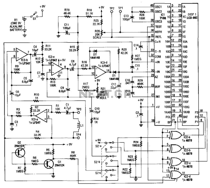
Using a pickup coil to drive an amplifier (IC3A-B-C-D), this meter circuit can be directly calibrated in field intensity units. R3/C3 and R12/C7 establish a frequency roll-off that compensates for the pickup coil's sensitivity and set a 20 kHz...
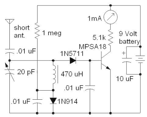
The following circuit illustrates a Field Strength Meter Circuit Diagram. Features: it is useful for radio frequency hobbyists and enthusiasts, and it uses only... The Field Strength Meter circuit is designed to measure the strength of radio frequency (RF) signals...
Figure 1 illustrates the schematic for a universal input, 7.6 V, 700 mA constant voltage/constant current (CV/CC) power supply designed for LED driver applications. This design employs the LinkSwitch-II product LNK606PG in a flyback configuration. The LNK606PG (U1) integrates...

Referring to the simplified schematic in A, the audio frequency generator (AFG) consists of 10 relatively simple circuit elements. IC1-c and IC1-d are configured as unity-gain non-inverting buffer amplifiers. The summing amplifier, IC2-c, combines equal amounts of the left...

A three-dimensional surround sound Ambisonic recording can be captured using a tetrahedrally arranged quartet of cardioid pattern microphone capsules connected to some simple circuitry to convert the outputs to a standard B-format signal. B-format signals represent a 3D soundfield...

This circuit serves as an alarm system suitable for both home security and personal belongings such as handbags. When installed in a home, it can be positioned on doors or windows, and when used for bags, it provides a...